
HUMAN EYE
Anatomy of the human eye
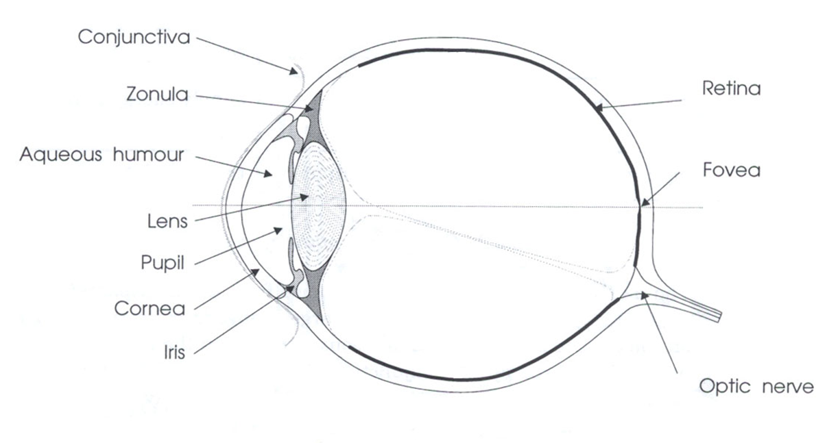
The eye allows us to see and interpret the shapes, colors, and dimensions of objects in the world by processing the light they reflect or emit. The eye is able to detect bright light or dim light, but it cannot sense objects when light is absent.
Light waves from an object enter the eye first through the cornea, which is the clear dome at the front of the eye. The light then progresses through the pupil, the circular opening in the center of the colored iris.
Fluctuations in incoming light change the size of the eye’s pupil. When the light entering the eye is bright enough, the pupil will constrict (get smaller), due to the pupillary light response.
Initially, the light waves are bent or converged first by the cornea, and then further by the crystalline lens (located immediately behind the iris and the pupil), to a nodal point (N) located immediately behind the back surface of the lens. At that point, the image becomes reversed (turned backwards) and inverted (turned upside-down).
The light continues through the vitreous humor, the clear gel that makes up about 80% of the eye’s volume, and then, ideally, back to a clear focus on the retina, behind the vitreous. The small central area of the retina is the macula, which provides the best vision of any location in the retina. If the eye is considered to be a type of camera (albeit, an extremely complex one), the retina is equivalent to the film inside of the camera, registering the tiny photons of light interacting with it.
Within the layers of the retina, light impulses are changed into electrical signals. Then they are sent through the optic nerve, along the visual pathway, to the occipital cortex at the posterior (back) of the brain. Here, the electrical signals are interpreted or “seen” by the brain as a visual image.
 Cornea
The cornea is the transparent, curved membrane on the front of the eye which serves as the outer window of the eye. The cornea is the primary (most powerful) structure focusing light entering the eye (along with the secondary focusing structure, the crystalline lens).
The cornea is composed, for the most part, of connective tissue with a thin layer of epithelium on the surface. Epithelium is the type of tissue that covers all free body surfaces.
The cornea is composed of 5 layers, from the front to the back:
- epithelium,
- Bowman’s (anterior limiting) membrane,
- stroma (substantia propria),
- Descemet’s (posterior limiting) membrane, and
- endothelium (posterior epithelium).
The transparency of the cornea is due to the fact that it contains hardly any cells and no blood vessels. However, blood vessels can creep in from around it, if it is constantly irritated or infected, which can interfere with vision.
On the other hand, the cornea contains the highest concentration of nerve fibers of any body structure, making it extremely sensitive to pain. The nerve fibers enter on the margins of the cornea and radiate toward the center. These fibers are associated with numerous pain receptors that have a very low threshold. Cold receptors also are abundant in the cornea, although heat and touch receptors seem to be lacking.
Sclera
Along its circumference, the cornea is continuous with the sclera: the white, opaque portion of the eye. The sclera makes up the back five-sixths of the eye’s outer layer. It provides protection and serves as an attachment for the extraocular muscles, which move the eye.
Tears and tear glandsCoating the outer surface of the cornea is a “pre-corneal tear film.” People normally blink the eyelids of their eyes about every six seconds to replenish the tear film. Tears have four main functions on the eye:
- wetting the corneal epithelium, thereby preventing it from being damaged due to dryness,
- creating a smooth optical surface on the front of the microscopically irregular corneal surface,
- acting as the main supplier of oxygen and other nutrients to the cornea,
- containing an enzyme called “lysozyme,” which destroys bacteria and prevents the growth of microcysts on the cornea, and
- flushing harmful bacteria and other microbes away from the eye, into the lacrimal canals and then out through the nose.
Iris
The iris, visible through the clear cornea as the colored disc inside of the eye, is a thin diaphragm composed mostly of connective tissue and smooth muscle fibers. It is situated between the cornea and the crystalline lens. The color(s), texture, and patterns of each person’s iris are as unique as a fingerprint.
The iris is composed of 3 layers, from the front to the back:
- endothelium,
- stroma, and
- epithelium.
The iris divides the anterior compartment, the space separating the cornea and the lens, into 2 chambers:
- the larger anterior chamber (between the cornea and the iris), and
- the smaller posterior chamber (between the iris and the lens).
 The color of the iris, established genetically, is determined by the amount of pigment present in the iris structure. No pigment at all (in the case of an albino) results in a pink iris. Some pigment causes the iris to appear blue. Increasing amounts of iris pigment produce green, hazel, and brown irises (or irides).
There actually are two pigments, melanin and lipochrome, which determine eye color. Melanin (brown) deposition is controlled by a gene on chromosome 15. Lipochrome (yellowish-brown) deposition is controlled by a gene on chromosome 19.
PupilIn the middle of a normal iris is the pupil. It is an opening that, typically, is circular and is comparable to the aperture of a camera. The pupil helps regulate the amount of light passing through to the retina, which is at the back of the eye.
As the amount of light entering the eye diminishes (such as in a dark room or at night), the iris dilator muscle (which runs radially through the iris like spokes on a wheel) pulls away from the center, causing the pupil to “dilate.” This allows more light to reach the retina. When too much light is entering the eye, the iris sphincter muscle (which encircles the pupil) pulls toward the center, causing the pupil to “constrict” and allowing less light to reach the retina. Certain eye drops also can dilate or constrict the pupils.
Crystalline lens
The transparent crystalline lens of the eye is located immediately behind the iris. It is composed of fibers that come from epithelial (hormone-producing) cells. In fact, the cytoplasm of these cells makes up the transparent substance of the lens.
The crystalline lens is composed of 4 layers, from the surface to the center:
- capsule,
- subcapsular epithelium,
- cortex, and
- nucleus.
The lens capsule is a clear, membrane-like structure that is quite elastic, a quality that keeps it under constant tension. As a result, the lens naturally tends towards a rounder or more globular configuration, a shape it must assume for the eye to focus at a near distance.
Slender but very strong suspensory ligaments, also known as zonules or zonules of Zinn, attach at one end to the lens capsule and at the other end to the ciliary processes of the circular ciliary body, around the inside of the eye. These thin ligaments or zonules hold the lens in place.
accommodationThe ciliary body is circular, and the ciliary muscle within it is a sphincter muscle, shaped like a tiny doughnut. The inside diameter of the muscle gets smaller when it contracts and larger when it relaxes.
When the eye is viewing an object at a far distance (such that parallel rays of light are entering the eye), the ciliary muscle within the ciliary body relaxes. The ciliary processes pull on the suspensory ligaments (or zonules), which in turn pull on the lens capsule around its equator.  This causes the entire lens to flatten or to become less convex, enabling the lens to focus light from the far-away object.
Conversely, when the eye views an object at a near distance, an “accommodative demand” is created. As a result, the ciliary muscle works or contracts, causing tension to be released on the suspensory ligaments and, subsequently, on the lens capsule. This causes both (front and back) lens surfaces to become more convex and the eye to be able to focus at near.
This adjustment in lens shape, to focus at various distances, is referred to as “accommodation” or the “accommodative process” and is associated with a concurrent constriction (decrease in size) of the pupil. The animated diagram above illustrates the change in stance of the ciliary body, crystalline lens, and pupil as the eye looks back and forth between far and near.
The “amplitude of accommodation” of an eye is the maximum amount that the eye’s crystalline lens can accommodate (change shape), in diopters (D). This amount is very high when a person is young and decreases with age.
The amplitude of accommodation is equivalent to the inverse (reciprocal) of the distance (“nearpoint of accommodation”) at which the emmetropic eye can focus clearly. (“Emmetropia” refers to an eye having no refractive error - no hyperopia, myopia, nor astigmatism - or it can refer to the optical system of an eye corrected to “plano” [0.00 diopters of refractive error] with glasses, contact lenses, or refractive surgery.)
Vision is a complicated process that requires numerous components of the human eye and brain to work together. The initial step of this fascinating and powerful sense is carried out in the retina of the eye. Specifically, the photoreceptor neurons (called photoreceptors) in the retina collect the light and send signals to a network of neurons that then generate electrical impulses that go to the brain. The brain then processes those impulses and gives information about what we are seeing. In this unit we will investigate the initial steps in the process of vision. We will discover how the photoreceptors work, and will specifically examine at the photoreceptor proteins to learn how light energy is converted into electrical energy. Additionally, we will examine some of the current studies that are helping to further our understanding of the proteins involved in the vision process.
Eye Anatomy and Function
Human anatomy has been studied since ancient times. For over 1400 years our understanding of anatomy was based on theories of the Greek physician, Galen of Pergamum (130-200 AD). However an accurate and comprehensive understanding of human anatomy was delayed until the Renaissance period, primarily because dissections and autopsies were forbidden by most religions. One of the first systematic studies of human anatomy which involved actual examination and dissection of the human body, was carried out by Andreas Vesalius (1514-1564). As a result of his extensive work, many of the previous misconceptions of Galenic medicine were corrected. The accumulated research of scientists over many hundreds of years has led to an excellent understanding of human anatomy .
A realistic understanding of the function of the components of the eye began around the 17th century, after the gross anatomy of the eye had been firmly established. It was realized in the 17th century that the retina, not the cornea as was previously thought, was responsible for the detection of light. Johannes Kepler of Germany and Renee Descartes of France, both prominent physicists of their time, made many advances in understanding vision. Much of their work applied the physical concepts of light rays and geometric optics to the vision process. Kepler first proposed that the lens of the eye focuses images onto the retina. A few decades later Descartes demonstrated that Kepler was correct. In a landmark experiment, Descartes surgically removed an eye from an ox and scraped the back of the eye to make it transparent. He then placed the eye on a window ledge as if the ox were looking out of the window. He looked at the back of the eye he and saw an inverted image of the scenery outside! Descartes correctly postulated that the image was inverted as a result of being focused onto the retina by the eye's lens.
Around the beginning of the 19th century Thomas Young, a prominent physicist and physician, carried out a number of studies on the eye that resulted in an understanding of how the lens focuses images onto the retina. He also showed that astigmatism results from an improperly curved cornea. We now understand that a number of vision disorders, including both near- and far-sightedness, also result from an improperly curved cornea. The lenses in eyeglasses function by correcting for the improper corneal curve.
We now know the basic function of the components of the human eye and how they participate in the vision process. Light that reflects off of objects around us is imaged onto the retina by the lens. The retina, which consists of three layers of neurons (photoreceptor, bipolar and ganglion) is responsible for detecting the light from these images and then causing impulses to be sent to the brain along the optic nerve. The brain decodes these images into information that we know as vision.

Microscopic Anatomy: Rod Cells and Cone Cells of the Retina
Although the microscope was first used in scientific observation in the late 16th and early 17th centuries, both the tool and the techniques of its use reached a sufficient level of sophistication by the 19th century to make it invaluable in examination of the structures of the eye. It was in the 1830's that several German scientists used the microscope to closely examine the retina. During this time that two different cells were discovered in the retina, the rod cells and cone cells. These cells were named because of their shape as viewed in the microscope.
A microscopic view of the rod cells of a zebrafish shows us how these cells actually look in an animal. Additional research showed that the rod and cone cells were responsive to light. Max Schultze (1825-1874) discovered that the retinal cones are the color receptors of the eye and the retinal rod cells while not sensitive to color, are very sensitive to light at low levels. Selig Hecht showed, in 1938, the exquisite sensitivity of rod cells when he showed that a single photon can initiate a response in a rod cell. Cone cells on the other hand are less sensitive to light but show great sensitivity to different colors. It is the cone cells that allow us to see in color. It is because cone cells remain unstimulated in low light environments that we do not see color in dimly lit places. Try this for yourself. Go into a closet and decrease the light level. Soon you will see only shades of gray. Slowly increase the light levels until you can begin to see color. This demonstration usually works well in a closet because of the many different colors of your clothes.
In the human eye, there are many more rod cells in the retina than there are cone cells. The number of rod cells and cone cells in animals is often related to the animal's instincts and habits. For example, birds such as hawks have a significantly higher number of cones than do humans. This let them to see small animals from a long distance away, allowing them to hunt for food. Nocturnal animals, on the other hand, have relatively higher numbers of rod cells to allow them better night vision.
A schematic drawing of rod and cone cells are shown in Figure 2. The cells are divided into two sections. The bottom portion is called the inner segment. It contains the nucleus and the synaptic ending. The synaptic ending attaches to the neurons which produce signals that go to the brain. The top portion is called the outer segment. The outer segment is comprised of a membrane which is folded into several layers of disks. The disks are comprised of cells that contain the molecules that absorb the light.
Visual Pigments
During the 1800's the visual pigments were discovered in the retina. Scientists, working by candlelight, dissected the retinas from frog eyes. When the retinas were exposed to day light they changed color. These scientists had discovered that the retina is photosensitive. They realized that the color they were observing was due to presence of a visual pigment, which was given the name rhodopsin. Later studies showed that rhodopsin is a protein that is found in the disks of the rod cell membrane.
Pigments are also found in cone cells. There are three types of cone cells, each of which contains a visual pigment. These pigments are called the red, blue or green visual pigment. The cone cells detect the primary colors, and the brain mixes these colors in seemingly infinitely variable proportions so that we can perceive a wide range of colors. Prolonged exposure to colors, for example when staring at a particular object, can cause fatigue in cone cells. This results in a change in the way that you perceive the color of the object that you are viewing. You will find a demonstration of the color fatigue effect on the Exploratorium's "Bird in a Cage" Web page.
The original theory of color vision was introduced by Thomas Young around 1790, prior to the discovery of the cone cells in the retina. Young was the first to propose that the human eye sees only the three primary colors, red, blue and yellow and that all of the other visible colors are combinations of these. It is now known that color vision is more complicated than this, but Young's work formed the foundation of color vision theory for the scientists that followed. The photoreceptor proteins of the cone cells have not yet been isolated. This may possibly be due to the difficulty in obtaining them. There are many fewer cone cells than rod cells in the retina. Also many animals do not have cone cells and hence do not see in color.
An Important Protein in the Rod Cell: Rhodopsin
George Wald and his coworkers at Harvard University pioneered our understanding of the molecules responsible for the first steps in the vision process. For this and other work on vision he was the recipient of the 1967 Nobel Prize in Medicine and Physiology. Wald's group was the first to elucidate the molecular components of the rod cell's functional protein rhodopsin. Prior to his work, rhodopsin was thought to be a chunk of molecular material. Wald and his co-workers determined that the protein consists of two molecular parts: a colorless amino acid sequence called opsin and a yellow organic chromophore called retinal.
It is now known that the rhodopsin protein has a molecular weight of ~40 kDa. The protein spans the membrane of the rod cell, and is therefore called a trans-membrane protein. The exact structure of rhodopsin has never been determined, however experimental data lead scientist to predict that it contains seven helices or turns. A schematic drawing of rhodopsin in the rod cell membrane is shown in Figure 3. About half of the protein is contained within the membrane with approximately 25% of the protein lying both above and below the membrane.
It is the rhodopsin protein in the retina that absorbs the light that enters the eye. Specifically, it is known that the retinal molecule, which is embedded inside rhodopsin, undergoes photo-excitation by absorbing light. In the photo-excitation process, the rhodopsin absorbs light and is excited to a higher electronic state. Numerous studies have been carried out to try to understand what happens after the rhodopsin absorbs light. Research has shown that upon photo-excitation the retinal part of rhodopsin undergoes a twisting around one of its double bonds (see Figure 4). The retinal then dissociates from the opsin. The change in geometry initiates a series of events that eventually cause electrical impulses to be sent to the brain along the optic nerve. Further research is needed to fully understand this complex process.
Vitamin A and Retinal
During the early part of the 20th century work continued on the frontier of research aimed at understanding vision. It was also around this time that the relationship between vision and proper nutrition began being studied at universities and agricultural schools. It had been shown during World War I that a vitamin A deficiency caused night blindness. The link between vitamin A and night blindness, however, did not become clear until George Wald and his coworkers isolated vitamin A from the retina in 1933. Prior to this finding the importance of vitamins was poorly understood. Additionally, the complete role of vitamins in physiological processes was unknown.
It is now understood that the human body makes retinal from vitamin A. A picture of retinal and vitamin A is shown in Figure 5. Both the retinal and vitamin A molecules contain a long chain of double bonds. When retinal dissociates from opsin, some of the retinal is destroyed. To replenish the destroyed retinal, it is important to have a source of vitamin A in your diet. Without this source of vitamin A, night blindness can develop as the rods can not function effectively without sufficient sources of retinal.